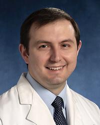
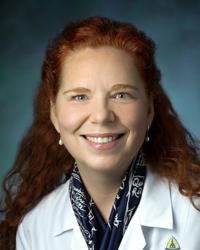
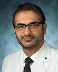
Stefan Zimmerman, MD
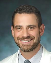
- Division Director, Diagnostic Imaging
- Professor of Radiology and Radiological Science
We offer state-of-the-art patient services in thoracic, abdominal and pelvic computed tomography (CT scan) and serve as a leading resource for education and research based on world-class CT imaging.
More than 90,000 CT examinations are performed each year. Our clinical service includes CT imaging of the chest, abdomen and pelvis, including CT angiography and 3D imaging. We have the latest in 3D software and currently complete more than 1,000 3D CT cases a month.
The section has nine state-of-the-art Siemens multidetector CT (MDCT) scanners (two scanners at MDCT 64 slice and seven flash dual source scanners). High temporal resolution results from the use of dual X-ray and detector systems. In flash mode an examination of the chest, abdomen and pelvis can be completed in 10-12 seconds.
The flash scanner is especially beneficial for patients who may experience low back pain or shortness of breath when lying down or patients who may have high or irregular heart rates.
Visit CTisus, the premier educational site for CT developed by Elliot K. Fishman, M.D.
The Division of Magnetic Resonance Imaging is committed to upholding the tripartite mission of Johns Hopkins Medicine, which is to offer its patients the highest-quality patient care, research and education available. The department’s world-renowned faculty focuses on blending the latest in magnetic resonance technology with the highest possible standard of patient care.
We have a team of highly specified experts in all aspects of body MRI, including hepatobiliary, pancreatic, cardiac, MR angiography, gastrointestinal, genitourinary, female pelvis and prostate MR. We provide multidisciplinary collaboration with other services, including internal medicine, cardiology, hepatology, urology, oncology and surgery, including transplantation. Referrals are welcome from physicians who do not have privileges at Johns Hopkins, as well as Johns Hopkins medical staff.
In the Body MRI section, we interpret over 7,000 body MRI examinations each year. MRI studies are performed on 14 state-of-the-art 1.5T and 3T in house clinical scanners, one interventional scanner and one intraoperative scanner in the IMRIS suite. In addition, we interpret body MRI examination from 5 offsite 1.5T and 3T magnets. We offer extensive clinical and research experience in all aspects of body MRI:
Cutting-edge research is actively ongoing in clinical body MRI in collaboration with other specialties. Research fellowships are available for one or more years. If you are interested in a research position in body MRI, please fill the online application form.
Novel research projects include
We offer state-of-the-art patient services in ultrasound and serve as a leading resource for education and research based on ultrasound imaging.
The ultrasound section of The Johns Hopkins Hospital performs more than 24,000 examinations each year, including abdominal and gynecologic ultrasound; thyroid and neck; obstetrics; organ transplants; ultrasound of various joints, muscles, tendons and nerves; carotid duplex, abdominal Doppler; and peripheral vascular Doppler examinations.
Our 15 rooms are equipped with state-of-the-art ultrasound units, including Philips Epiq and IU22, Siemens S3000 Helix and S2000, and GE Logiq E9. Most of our units have 3-D and fusion capabilities. We currently store all images electronically on a picture archiving and communicating system.
Our sonographers are registered by the American Registry of Diagnostic Medical Sonographers, and our facilities are accredited in abdominal, obstetrical, gynecological and vascular sonography by the American College of Radiology.
Diagnostic Radiology Residency
Training at Johns Hopkins Medicine is a truly unique experience. As a world-renowned referral center with over a thousand beds, the breadth and volume of pathology are simply unmatched. Our philosophy is to allow residents to increasingly build responsibility and decision-making capabilities from learning the very basics in the first year to becoming the radiology point person during overnight call.
Body MRI Fellowship
The dedicated Body MRI fellowship is an intensive 12-month clinical fellowship in Body MRI including chest, cardiac, vascular, abdomen, hepatobiliary, pelvis, prostate and musculoskeletal MRI studies.
Cardiothoracic Imaging Fellowship
The Cardiothoracic Imaging fellowship program will accept one fellow each year and will be fully integrated with Johns Hopkins Hospital. The fellow will rotate through both hospitals to take advantage of the unique and complementary educational opportunities as well as the exceptional resources that are available across the Hopkins Health System.
Cross-Sectional Body Imaging Fellowship
The primary mission of the fellowship is to provide superior clinical training in advanced CT, ultrasound and MR imaging of the body. This mission is accomplished by the dedication of faculty members who are deeply committed to resident and fellow education.

