-
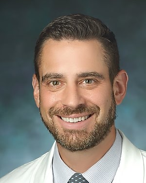
Stefan Zimmerman, MD

- Division Director, Diagnostic Imaging
- Professor of Radiology and Radiological Science
CARDIAL - Cardiovascular Research, Diagnostic Imaging, and AI Lab
At CARDIAL, our mission is to pioneer advancements in cardiovascular imaging through relentless innovation and a commitment to scientific integrity. Our goal is to enhance diagnosis, guide treatment, and improve prognosis for cardiovascular diseases, ultimately contributing to better patient outcomes and a healthier world.
We are dedicated to training the next generation of researchers, equipping them with the skills and knowledge to lead future breakthroughs. We embrace diversity and inclusion, recognizing that a variety of perspectives and backgrounds enrich our research and drive creative solutions. We are committed to fostering an environment where all individuals are valued, respected, and empowered to achieve their full potential.
Research Focus
- Cardiac Imaging/ Volumetric and Functional Assessment
- Vascular and Aortic Imaging by MRI and CT
- Myocardial Tissue Characterization by MRI and CT
- Myocardial Deformation Analysis by MRI and CT
- Hemodynamics using 4-Dimensional Flow MRI
- Computational Fluid Dynamics
- Population Imaging
- Radiomics analysis of Images
- Artificial Intelligence and Statistical Machine Learning
- Deep Learning-based Image analysis
Lab Members
Post-doctoral Fellows:
- Ghazal Zandieh
- Shadi Afyouni
Students:
- Emma Enriquez
- Justin Chiuwei
- Winston Wongso
- Ronit Sohal
- Karunya Chittamuri
Alumni:
- Ali Borhani (radiology resident at JHH)
- Iman Yazdani Nia (fellow at Upenn, Philadelphia)
Current Projects
- Myocarditis/Myositis: This project aims to use cardiac MRI to tackle major gaps and unmet clinical needs in the prospective, systematic detection of myocarditis in Idiopathic inflammatory myopathies (IIM) and the risk stratification of these patients with myocarditis to predict the likelihood of mortality and or cardiac endpoints such as heart failure. Chief Collaborator: Dr. Julie Paik.
- Amyloidosis: This project aims to use cardiac MRI to accurately diagnose cardiac amyloidosis through findings such as abnormal myocardial null time on inversion recovery scout, diffuse subendocardial late gadolinium enhancement, increased native T1 time, and expanded extracellular volume. In addition, the goals are to identify the prognostic utility of cardiac MRI parameters in amyloidosis patients, in conjunction with other markers from nuclear medicine techniques, clinical history, and biopsies. Chief Collaborator: Dr. Joban Vaishnav.
- Cardiac MRI in non-ischemic cardiomyopathy (NICM): The primary goals of this project are to develop and evaluate a model based on cardiac MRI parametric mapping that can identify NICM phenotypes. This also involves incorporating a variety of radiomics features into a discriminative model for NICM. We also aim to construct and validate models using the same cardiac MRI parametric mapping features designed to forecast disease progression and outcomes in patients with NICM. Chief Collaborator: Dr. Jose Madrazo.
- Myocardial Fat: This study seeks to explore the association between myocardial tissue heterogeneity derived from both non-contrast and contrast-enhanced cardiac CT and adverse cardiovascular events including ventricular arrhythmia occurrences using a combination of machine learning and radiomics techniques. Chief Collaborators: Drs. Jonathan Chrispin, Katherine Wu, and Joao Lima.
- Connective Tissue Disorders/Aortic Imaging: The primary goal of this project is to combine artificial intelligence-enabled image processing and analytics, advanced imaging, and computational fluid dynamics-based simulations to create a Digital Twin of the aorta and other vascular structures. Digital Twin technologies can be potentially useful for risk assessment and continuous strategic management of patients with vascular connective tissue disorders. Chief Collaborators: Drs. Hal Dietz, Tony Guerrerio, and Paul Burke.
- Pulmonary Hypertension/Systemic Sclerosis: The primary goal of this project is to phenotype patients with systemic sclerosis, idiopathic pulmonary hypertension, and those at the intersection of the two conditions using cardiac MRI, as a means to more effectively assess risk and improve patient management. We study the role of cardiac MRI in conjunction with echocardiography, right heart catheterization, and other clinical features. Chief Collaborators: Drs. Paul Hassoun, Monica Mukherjee, and Steve Hsu.
- Multiple Cardiac Imaging Projects that involve optimization of protocols, procedures and diagnosis techniques. Chief Collaborators: Drs. Claire Brookmeyer and Muhammad Umair

