-
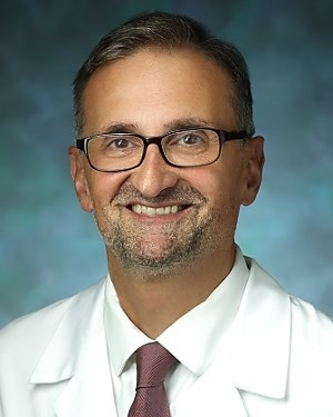
Armin Arbab-Zadeh, MD MPH

- Director, Cardiac Computed Tomography
- Professor of Medicine
Primary Location: The Johns Hopkins Hospital, Baltimore, MD
-
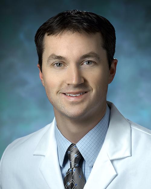
Michael Joseph Blaha, MD MPH

- Director of Clinical Research, Ciccarone Center for the Prevention of Cardiovascular Disease
- Professor of Medicine
Primary Location: Johns Hopkins Health Care & Surgery Center - Green Spring Station, Lutherville, Lutherville, MD
-
Henry R. Halperin, MD

- David J. Carver Professor in Medicine
- Professor of Medicine
Primary Location: The Johns Hopkins Hospital, Baltimore, MD
-
Allison Hays, MD

- Medical Director of Echocardiography Programs of the Johns Hopkins Hospital
- Associate Professor of Medicine
Primary Location: Johns Hopkins Bayview Medical Center, Baltimore, MD
-
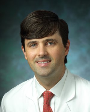
AV Kolandaivelu, MD

- Assistant Professor of Medicine
Primary Location: Johns Hopkins Health Care & Surgery Center - Green Spring Station, Lutherville, Lutherville, MD
-
Joao Lima, MD

- Director of Cardiovascular Imaging
- Professor of Medicine
Primary Location: The Johns Hopkins Hospital, Baltimore, MD
-
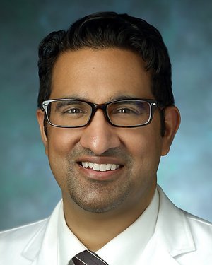
Raj Mukherjee, MD MPH

- Director of Neurosurgical Oncology, Johns Hopkins Bayview Medical Center
- Associate Professor of Neurological Surgery
Primary Location: Johns Hopkins Bayview Medical Center, Baltimore, MD
-
Seamus Paul Whelton, MD MPH

- Director of the Advanced Subclinical Atherosclerosis Clinic
- Associate Professor of Medicine
Primary Location: Johns Hopkins Health Care & Surgery Center - Green Spring Station, Lutherville, Lutherville, MD
-
Katherine Wu, MD

- Associate Professor of Medicine
Primary Location: The Johns Hopkins Hospital, Baltimore, MD
Imaging
Johns Hopkins researchers are working to develop improved imaging of the heart, which is challenging considering its constant motion. Imaging is a noninvasive way to tell doctors a lot about the structure and functioning of a patient’s heart.
Imaging for Catheter Ablations
One active area of research is how noninvasive imaging can guide catheter ablation procedures that modify electrical circuits in the heart to treat arrhythmias or irregular heartbeats. Some of the imaging techniques include computed tomography using X-rays, MRI and ultrasound to guide a physician when performing a cardiac ablation. During a cardiac ablation, a catheter is moved into the heart and used to destroy small regions of the heart wall, which may be making the heart’s rhythm irregular.
Imaging to Diagnose Heart Dysfunction at an Early Stage
Researchers are looking at ways to improve early detection of heart dysfunction to help prevent serious heart problems that result from a weak heart, such as heart failure. Specialized imaging techniques such as speckle tracking echocardiography and strain MRI are powerful noninvasive methods to help detect early heart dysfunction and allow rapid treatment to improve patient outcomes. These techniques are especially useful in monitoring heart function with certain chemotherapy drugs, which are used to treat cancer but have potentially harmful effects on the heart.
Imaging to Help Define Risk and Diagnose Coronary Artery Disease
With advanced imaging, physicians can search for clues that someone is at risk for heart attack or other cardiac problems. Separating patients into higher risk and lower risk categories can help researchers create benchmarks for treatment. In patients with coronary artery disease, the results of imaging tests can help determine which medication or procedure is best for a specific patient.
Safe MRI Techniques for Patients with Pacemakers
Traditionally, MRIs and implanted cardiac devices, such as pacemakers, did not mix. The powerful magnetic and radiofrequency fields could potentially cause the device to malfunction. After 15 years of research and trials, Johns Hopkins researchers developed protocol for patients with implanted devices. They can reprogram pacemakers to a safe mode while a patient has an MRI scan. They carefully monitor the patient’s blood pressure, electrical activity of the heart and oxygen saturation, and look for any unusual symptoms throughout the process. The protocol continues to be fine-tuned as more research is done. Making MRIs more available to patients with pacemakers is important for those who could benefit from early detection of cancer and other diseases, as well as for guiding surgeons during procedures.
Dr. Joao Lima
Dr. Lima is a cardiologist whose clinical practice focuses on the use of cardiac imaging, such as echocardiography, CT scans and MRI, to access a patients risk for heart attack, stroke and heart failure.
Related Articles & Press Releases
- A Clearer Picture of the Heart?
- ‘High-Normal’ Blood Pressure in Young Adults Spells Risk of Heart Failure in Later Life
- The Newest CT: Faster Than a Heartbeat
- Squeeze and show
- Hopkins Study Finds MRI Tests Safe for People with Implanted Cardiac Devices When Certain Guidelines are Followed
- Safety guidelines for MRI with ICD
- Updated Classification System Captures Many More People at Risk for Heart Attack


