Research Lab Results
-
Cardio-Obstetrics Research
Under the division of Maternal-Fetal Medicine, our Cardio-Obstetric research efforts seek to advance the field of gynecology through medical care and innovation. With a focus on the effect of heart conditions on pregnancy and the ways in which pregnancy can put stress on your heart and circulatory system, our goal in this multi-disciplinary research is that our findings may lead to the development of new treatments or preventative therapies for patients and their babies to better manage a heart condition during pregnancy. -
Clare Rock Lab
Dr. Clare Rock is an assistant Professor of Medicine, Division of Infectious Diseases at the Johns Hopkins University School of Medicine, Associate hospital Epidemiologist at the Johns Hopkins Hospital, and Faculty Member at Armstrong Institute for Patient Safety and Quality. Her research interest focuses the prevention of pathogen transmission in the hospital environment. This includes novel strategies of improving patient room cleaning and disinfection, including human factors engineering approaches, and conducting robust clinical trials to examine effectiveness of ""no touch"" novel technologies such as UV-C light. She has particular interest in carbapenem-resistant Enterobacteriaceae transmission in the hospital environment, including outbreak management, and transmission and epidemiology of Clostridium difficile. Her other area of interest is diagnostic stewardship, and the behavioral, cultural and human factors aspects of implementation of initiatives to enhance appropriate use of diagnostic tests. She leads a national initiative, as part of the High Value Practice Academic Alliance, examining strategies for appropriate testing for Clostridium difficile. This is a wider implementation of work that Dr. Rock conducted with The Johns Hopkins Health System facilities. Dr. Rock has multiple sources of grant funding including from the Agency of Healthcare Research and Quality, Centers for Disease Control and Prevention, and industry. Dr. Rock is Vice Chair of the Society for Healthcare Epidemiology of America Research Network, and serves on the SHEA research committee. Dr. Rock earned her M.B.B.Ch. at the University College Dublin School of Medicine, National University of Ireland, and her MS masters of clinical science of research at the University of Maryland, where she received the MS scholar award for epidemiology. -
Cammarato Lab
The Cammarato Lab is located in the Division of Cardiology in the Department of Medicine at the Johns Hopkins University School of Medicine. We are interested in basic mechanisms of striated muscle biology. We employ an array of imaging techniques to study “structural physiology” of cardiac and skeletal muscle. Drosophila melanogaster, the fruit fly, expresses both forms of striated muscle and benefits greatly from powerful genetic tools. We investigate conserved myopathic (muscle disease) processes and perform hierarchical and integrative analysis of muscle function from the level of single molecules and macromolecular complexes through the level of the tissue itself. Anthony Ross Cammarato, MD, is an assistant professor of medicine in the Cardiology Department. He studies the identification and manipulation of age- and mutation-dependent modifiers of cardiac function, hierarchical modeling and imaging of contractile machinery, integrative analysis of striated muscle performance and myopathic processes. -
Zackary Berger Lab
The research mission of the Zackary Berger Lab is to bridge evidence-based medicine and shared decision-making in the context of patient-centered care. Lab studies investigate how to accomplish this in the common case of uncertainty, while seeking to clarify the ethics of decision-making and empirically describe how shared decision-making is and should be done. Zackary Berger, MD, PhD, is an assistant professor in the Division of General Internal Medicine at the Johns Hopkins School of Medicine. In addition to his work as an internist and primary care physician, Dr. Berger is an associate faculty member in the Berman Institute of Bioethics, and core faculty in the Evidence Based Practice Center as well as the Center for Health Services and Outcomes Research.
-
Dmitri Artemov Lab
The Artemov lab is within the Division of Cancer Imaging Research in the Department of Radiology and Radiological Science. The lab focuses on 1) Use of advanced dynamic contrast enhanced-MRI and activated dual-contrast MRI to perform image-guided combination therapy of triple negative breast cancer and to assess therapeutic response. 2) Development of noninvasive MR markers of cell viability based on a dual-contrast technique that enables simultaneous tracking and monitoring of viability of transplanted stems cells in vivo. 3) Development of Tc-99m and Ga-68 angiogenic SPECT/PET tracers to image expression of VEGF receptors that are involved in tumor angiogenesis and can be important therapeutic targets. 4) Development of the concept of “click therapy” that combines advantages of multi-component targeting, bio-orthogonal conjugation and image guidance and preclinical validation in breast and prostate cancer models.
-
Xiao Group
The objective of the Xiao Group's research is to study the dynamics of cellular processes as they occur in real time at the single-molecule and single-cell level. The depth and breadth of our research requires an interdisciplinary approach, combining biological, biochemical and biophysical methods to address compelling biological problems quantitatively. We currently are focused on dynamics of the E. coli cell division complex assembly and the molecular mechanism in gene regulation. -
Wendy Bennett Lab
I am a primary care doctor and public health researcher committed to improving women’s health and health care across their lives and improving gender and racial equity.
I am an Associate Professor of Medicine in The Johns Hopkins University School of Medicine, Division of General Internal Medicine. My research focuses on identifying strategies to prevent and manage obesity and type 2 diabetes and cardiovascular disease, particularly among women at highest risk due to pregnancy complications. I conduct pragmatic and community-based randomized controlled trials to test high impact and scalable strategies to reduce excessive weight gain in pregnancy, reduce postpartum weight retention and cardiometabolic risk.
I hold several leadership positions, and I am the Director of Research at Johns Hopkins Community Physicians, the Co-Director of the Johns Hopkins Center for Women’s Health, Sex and Gender Research and a Core Faculty Member of the Welch Center for Prevention, Epidemiology and Clinical Research.
-
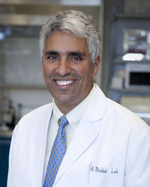
William Bishai Laboratory
The William Bishai Laboratory studies the molecular pathogenesis of tuberculosis. The overall goal of our laboratory is to better understand tuberculosis pathogenesis and then to employ this understanding toward improved drugs, vaccines and diagnostics. Since Mycobacterium tuberculosis senses and adapts to a wide array of conditions during the disease process, it is clear that the regulation of expression of virulence factors plays an important role in pathogenesis. As a result, a theme of our research is to assess mycobacterial genes important in gene regulation. We are also interested in cell division in mycobacteria and the pathogenesis of caseation and cavitation. -
Venu Raman Research Lab
The Raman laboratory is within the Division of Cancer Imaging Research in the Department of Radiology and Radiological Science. The focus of the laboratory is bench-to-bed side cancer research. We integrate molecular and cellular biology, developmental biology, cancer biology, molecular imaging techniques to study cancer formation and progression. Many of the projects in the lab investigate dysregulated genes in cancer and the translatability of this information to a clinical setting. One such project is to functionally decipher the role of a RNA helicase gene, DDX3, in the biogenesis of multiple cancer types such as breast, lung, brain, sarcoma, colorectal and prostate. Additionally, using a rational drug design approach, a small molecule inhibitor of DDX3 (RK-33) was synthesized and its potential for clinical translation is being investigated.
-
Vestibular NeuroEngineering Lab
Research in the Vestibular NeuroEngineering Lab (VNEL) focuses on restoring inner ear function through “bionic” electrical stimulation, inner ear gene therapy, and enhancing the central nervous system’s ability to learn ways to use sensory input from a damaged inner ear. VNEL research involves basic and applied neurophysiology, biomedical engineering, clinical investigation and population-based epidemiologic studies. We employ techniques including single-unit electrophysiologic recording; histologic examination; 3-D video-oculography and magnetic scleral search coil measurements of eye movements; microCT; micro MRI; and finite element analysis. Our research subjects include computer models, circuits, animals and humans. For more information about VNEL, click here. VNEL is currently recruiting subjects for two first-in-human clinical trials: 1) The MVI Multichannel Vestibular Implant Trial involves implantation of a “bionic” inner ear stimulator intended to partially restore sensation of head movement. Without that sensation, the brain’s image- and posture-stabilizing reflexes fail, so affected individuals suffer difficulty with blurry vision, unsteady walking, chronic dizziness, mental fogginess and a high risk of falling. Based on designs developed and tested successfully in animals over the past the past 15 years at VNEL, the system used in this trial is very similar to a cochlear implant (in fact, future versions could include cochlear electrodes for use in patients who also have hearing loss). Instead of a microphone and cochlear electrodes, it uses gyroscopes to sense head movement, and its electrodes are implanted in the vestibular labyrinth. For more information on the MVI trial, click here. 2) The CGF166 Inner Ear Gene Therapy Trial involves inner ear injection of a genetically engineered DNA sequence intended to restore hearing and balance sensation by creating new sensory cells (called “hair cells”). Performed at VNEL with the support of Novartis and through a collaboration with the University of Kansas and Columbia University, this is the world’s first trial of inner ear gene therapy in human subjects. Individuals with severe or profound hearing loss in both ears are invited to participate. For more information on the CGF166 trial, click here.