The Johns Hopkins Division of Maternal-Fetal Medicine is home to an exceptional group of maternal-fetal medicine specialists, ultrasonographers, nurse practitioners and genetic counselors, whose clinical mission is to provide comprehensive and specialized ultrasound services for both high-risk and low-risk pregnancies.
Our Services
- First-trimester scans including dating/viability, first trimester screening and nuchal translucency screening, detailed early anatomy, early echocardiogram and preeclampsia screening
- Detailed fetal anatomy screens
- Serial cervical screening for history of preterm delivery
- Fetal growth ultrasound
- Multiple gestations including TTTS monitoring
- Fetal cardiac arrhythmia evaluation and screening for congenital heart block
- Second opinion/consultation for known/suspected fetal abnormalities
- Fetal echocardiograms
- Neurosonograms
- Evaluation of placental abnormalities, including placenta previa and placental accreta spectrum disorder
Specialty Clinics
What to Expect at your OB Ultrasound
Get ready for your OB ultrasound appointment at Johns Hopkins Medicine. As part of our commitment to offering compassionate, comprehensive care for your pregnancy, this informative video covers what to expect and how to prepare.
Watch the Spanish version (en Espanol)Our Locations
-
Johns Hopkins Hospital
600 North Wolfe Street
Nelson Building, 2nd Floor
Baltimore, MD 21287
-
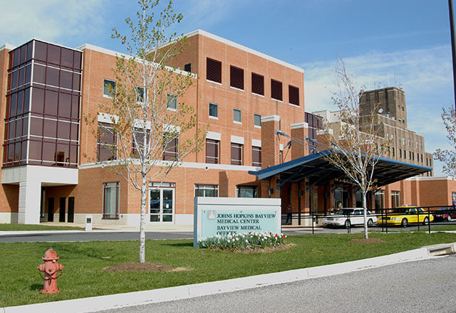
Bayview Medical Offices
4940 Eastern Avenue,
1st Floor, Clinic #2
Baltimore, MD, 21224
-
Johns Hopkins Perinatal Ultrasound at Green Spring Station
10755 Falls Road Pavilion I
Suite 470
Lutherville, MD 21093
-
Johns Hopkins Women's Services at White Marsh
4924 Campbell Blvd
Suite 225
White Marsh, MD 21236
-
Johns Hopkins Community Physicians - Canton Crossing
1501 S. Clinton Street
Suite 200
Baltimore, MD 21224
-
Johns Hopkins Community Physicians - Remington
2700 Remington Avenue
Suite 2000
Baltimore, MD 21211
Frequently Asked Questions
-
Ultrasound is a specialized exam using sound waves (not X-rays) to visualize your fetus. No radiation is involved. To learn more, please visit the American Institute of Ultrasound in Medicine (AIUM).
-
Obstetric ultrasound at Johns Hopkins is AIUM-accredited and employs registered ultrasonographers or diagnostic medical sonographer candidates who specialize in the field of obstetrics and high-risk obstetrics. All scans are interpreted by one of our maternal-fetal medicine specialists. In most cases, study results are provided to the patient before departure from our division.
-
- Please be sure to arrive at least 10 to 15 minutes prior to your scheduled appointment to allow time for registration.
- Please have your referral and a current insurance card with you at the time of your visit.
- Due to the size of some of our exam rooms and the concentration needed on behalf of your sonographer, and to ensure your privacy, we kindly ask that you limit your guests to two adults.
- If you arrive late for your appointment, we unfortunately cannot guarantee that we will be able to accommodate you the same day. If you know that you are going to be late, please call ahead at 410-955-8976 so that we may discuss accommodations with you.
-
The length of your exam may vary depending on the type of exam being performed, the number of fetuses and fetal cooperation. For a singleton pregnancy, you may expect 45 minutes to an hour, which includes chart review, exam time, and exam review by your physician and the sonographer. Twins and higher-order multiples are allotted the proper amount of time needed to perform a complete exam. Please note that if there is an unexpected finding, your exam may take longer, and if the fetus is uncooperative, you may need to return on a different day to try to complete the study.
-
You will need a full bladder if:
- You are less than 14 weeks pregnant.
- You are having a CVS procedure.
Your bladder should to be partially full if:
- You are between 14 to 28 weeks pregnant.
You do not need a full bladder if:
- You are having amniocentesis.
- You are more than 28 weeks pregnant.
-
It is fine to eat before your ultrasound, unless specifically instructed otherwise by a physician.
-
Digital recording for nondiagnostic purposes is not performed, but keepsake still images of your fetus may be provided. Please be aware that fetal position, gestational age and maternal build may limit our ability to obtain optimal keepsake images.
-
While we do have 3-D/4-D ultrasound machines, they are reserved for cases in which there is a known or suspected fetal abnormality.
In the event of a fetal abnormality, 3-D/4-D technology may sometimes be beneficial, but the limitations of 3-D are often the same as 2-D. Therefore, this technology is used at the attending physician’s discretion.
The Food and Drug Administration cautions against the use of 3-D/4-D imaging for entertainment purposes. Click here to read the official statement of the American Institute of Ultrasound in Medicine regarding the use of 3-D/4-D technology.
-
Depending upon gestational age, fetal position and maternal build, we may be able to tell you the sex we think your fetus will be, but please keep in mind that ultrasound is not 100 percent accurate in the fetal sex determination.
-
At 20 weeks of gestation, it is recommended that pregnant women have an ultrasound to confirm if the fetus is alive, measure growth, detect uterine or placental abnormalities, assess fluid volume, and image all fetal organ systems to detect any abnormalities.
-
At any point during your pregnancy or as routine practice, you may be asked to return after your 20-week anatomic survey to evaluate the growth of the fetus. We will evaluate the fluid volume, perform measurements of your fetus and image some major organ systems again if fetal position allows. We may also assess fetal blood flow when appropriate.
-
While most fetuses are structurally normal, some may have major or minor problems. In the event that we detect any abnormality, you will speak with a maternal-fetal medicine specialist prior to departure from our division. Please note that there are limitations to ultrasound, and not all fetal abnormalities can be detected prenatally.
-
The Johns Hopkins Hospital prides itself on the clinical education provided to our students, including diagnostic medical sonography students, medical students, residents and fellows in training. While we hope that you will take part in their education, we understand if you have reservations and respect your right to refuse. Regardless of your choice, the quality of your exam will not be impacted, and a trained, registered sonographer will thoroughly evaluate your fetus.

