Venous Malformation
What are VMs?
Veins are part of the circulatory system that moves blood through the body. Veins carry blood from the body back to the heart. The heart pumps the blood through the lungs so that it can pick up oxygen. The body uses oxygen to make energy. Venous malformations (VMs) occur when veins do not form normally. VMs can be completely isolated from normal veins or can drain into them. VMs are not part of the normal vein system.
VMs are the most common type of vascular anomaly. They can occur anywhere on the body, but are most common on the head and neck.
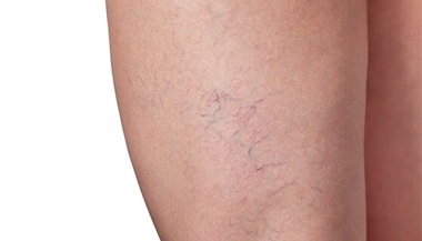
VMs can look like a bruise on the skin or a growth under the skin. VMs typically have a bluish color if in or near the skin.
In many cases, when a VM is found, it is already bigger than 5 centimeters (the size of a plum). When pressing down on a VM, it will often shrink, like a balloon losing air. After stopping, it will fill back up, like a balloon filling with air. This is the result of blood being pushed out of the malformation and then refilling slowly.
Round, hard spots can sometimes be felt when pressing on a VM. These are called phleboliths, which are calcified blood clots within the malformation. They are often about the size of a pearl.
Pain, swelling and disfigurement are the most common symptoms of VMs. Swelling or pain may come and go or may occur all of the time. Sometimes this may be due to a clot that forms within the malformation. A VM near a joint, such as an elbow or knee, can cause that joint to function badly. A VM near a nerve can lead to damage of that nerve and can cause pain or poor function.
Many people with visible VMs may feel embarrassed or self-conscious. Caregivers and teachers need to be aware to watch for teasing or bullying by classmates of a child with a VM.

Illustration of child with venous malformation on her face.
© Eleanor Bailey

Illustration of woman with venous malformation on her tongue.
© Eleanor Bailey

Illustration of venous malformation with macrocystic spaces, meaning with debris separated by septa.
© Eleanor Bailey
How are VMs diagnosed?
VMs are sometimes found deep in the body, and cannot be seen under the skin. VMs are sometimes found on an imaging study such as an X-ray or an MRI performed for other reasons, or because of symptoms, such as swelling or pain.
Most VMs are not hereditary. That is, they are not passed from parent to child. Nothing that a mother does during pregnancy can cause or prevent these malformations.
Some very rare kinds of VMs are hereditary. This means they are carried in the family’s DNA and can happen to other family members.
Most VMs can be diagnosed by a doctor who is skilled in caring for patients with them. This doctor will ask about the patient’s growth and development and do a physical examination.
An MRI is the best imaging study to diagnose a VM. It provides a detailed scan or picture of the inside of the body to help see the size and location of the VM. An MRI will also show other important structures, such as nerves, muscles, arteries and veins that are near the VM and that may be affected by treatment.
Magnetic resonance angiography (MRA) or magnetic resonance venography (MRV) are specific types of MRI that show blood vessels and blood flow. MRA/MRV can help to show whether there are arteries connecting to the VM or to see if there are veins that are draining blood from the VM.
MRI/MRV/MRA do not expose a patient to radiation. Images are created using powerful magnets.
An ultrasound is also useful to diagnose and monitor a VM. An ultrasound uses sound waves to make a picture of the blood vessels and tissues under the skin. Ultrasound can also be used to detect the speed of blood flow, which helps doctors diagnose a VM. It is a good method for young children because it doesn’t require a child to lie very still and because it does not expose a patient to radiation. Also, it can be done while a child is awake. However, ultrasound does not provide as much information about nearby anatomy as MRI does.
Occasionally, a doctor may do a CT scan to see whether a VM is affecting a bone. A CT scan is like an MRI, except it uses X-rays instead of magnets. In general, CT is not the best way to diagnose a VM
More Information on Vascular Anomalies from Johns Hopkins Medicine
Our Approach to Treating Vascular Anomalies
Johns Hopkins Medicine has developed a multidisciplinary Vascular Anomalies Center in order to offer patients individualized treatment. As a leader in diagnosing, researching and treating vascular anomalies and vascular tumors, our team of specialists provide comprehensive treatment and care.
How are VMs treated?
Most VMs grow as the patient grows. VMs can also grow after trauma or grow even faster during puberty or pregnancy. VMs are rarely cured and many patients will need to be treated at different times throughout their lives.
Treatment is typically focused on managing the VM to decrease the size and symptoms or to reduce or prevent problems that can be caused by the malformation. Although VMs cannot often be completely cured, there are many ways to manage them well.
VM is benign, which means it is not cancer. If a VM is not causing problems, such as pain or loss of function or deformity, active watchful waiting may be the best treatment. Once a VM starts causing problems, treatment will begin.
If a VM is in a sensitive or dangerous area, it may need treatment even if it has not yet begun to cause problems.
We recommend early evaluation by a specialist if you are concerned that you or your child may have a VM. Treatment is individualized. Your doctors will partner with you to make sure you or your child are getting the right treatment at the right time.
A team of doctors who specialize in treating vascular anomalies (tumors and malformations) will work together to treat a VM. The treatment team may include interventional radiologists, diagnostic radiologists, dermatologists, surgeons, hematologists and geneticists.
An interventional radiologist is a doctor who can read pictures and scans of the body and use these images to treat the VM without cutting the skin. Interventional radiologists play a central role in diagnosing and treating a VM.
A surgeon may be able to help correct disfigurement or deformity from VM, once most of the malformation has been treated. Large VMs can lead to problems with blood clotting.
A hematologist is a doctor who treats blood diseases and will make sure that blood is clotting properly before, during and after any procedures. Some VM and other vascular malformations can be treated with medications such as sirolimus, which require checking bloodwork and monitoring with a specialist who has experience using these medicines for patients with VM.
A dermatologist treats skin diseases. When a VM involves the skin, a dermatologist may be able to help treat involved skin with laser therapy.
A geneticist is a doctor who studies diseases that are passed on from parents to children through their genes. Geneticists help patients better understand their condition and discuss what risk, if any, exists for passing the risk of having a VM to future children.
Sclerotherapy to Treat VM
Sclerotherapy is a very useful treatment for VM and is performed by an interventional radiologist. In sclerotherapy, ultrasound is used to target the VM and X-ray imaging helps to guide and monitor the treatment.
The skin is not cut. While the patient is asleep, needles are used to deliver a liquid medicine, called a sclerosant, directly into the abnormal veins that make up the VM. This medicine damages and destroys the abnormal veins. Most sclerosants will cause the blood inside the VM to clot and will immediately damage the abnormal veins. Others have a more delayed effect. Either way, the goal of sclerotherapy is to cause the malformation to scar so that little or no blood will flow through the VM. This will cause the VM to shrink.
Several sclerotherapy treatments are often needed. Treatments are usually at least six weeks apart. Sclerotherapy makes the VM get smaller, but VMs can get bigger again over time. Typically VMs are managed throughout life, not cured. The goal of treatment is to improve symptoms as much as possible.
Preparing for Treatment
Before sclerotherapy, the treatment team will prepare you for what normally happens after the procedure and for any potential problems.
Most patients are put to sleep under general anesthesia during sclerotherapy. Some patients will be able to go home the day of the procedure, and some will stay overnight in the hospital to recover.
Following treatment, there could be swelling, irritation on the skin and bruising at the treatment site.
Ulceration is the most common complication of sclerotherapy. An ulcer is a sore or wound. Ulceration happens in less than 5 percent of cases. If an ulcer develops, your treatment team will treat it.
Additional Treatments for VMs
Sometimes laser therapy is used to treat VMs that affect the skin. On occasion surgery can help correct deformity or loss of function. For most vascular anomalies, a combination of treatment methods is best.
Doctors are working on new treatments for VMs. For extensive VMs, some patients can be treated with medications such as sirolimus. A medicine called sirolimus (rapamycin) has worked for some patients. Because sirolimus can suppress the immune system, close monitoring by an experienced specialist is necessary.
Surgery may be needed after sclerotherapy to remove a mass, extra skin or a deformity left by the VM. Small VMs can sometimes be treated with surgery alone. VMs will often return after surgery because it is very difficult to remove a VM completely. Only surgeons with experience in treating complicated vascular malformations should operate on them.
Other therapies, such as cryoablation (freezing therapy) and laser/radiofrequency ablation (heating therapy) are sometimes used to treat VM.
VMs that involve the skin are sometimes treated with different types of lasers, such as a pulsed-dye laser, a long-pulsed Nd:YAG laser or others. Several laser treatments are typically needed for best results for these skin-based VMs. These laser treatments are typically spaced 4 to 12 weeks apart.


