Glomuvenous Malformation
What You Need to Know
- Glomuvenous malformations are caused by mutations (changes) in genes that result in malformed veins.
- The malformed veins show up as bumps or marks under the skin, most commonly in the fingers and toes, but they may occur elsewhere in the body.
- In about 90% of cases, glomuvenous malformations are solitary, meaning the child has only one.
- Glomuvenous malformations can be removed with surgery or other treatments.
What is a glomuvenous malformation?
Glomuvenous malformations (GVMs) are misshapen veins that are present at birth. Usually, a child has one GVM (solitary), but multiple lesions are possible, and more lesions can emerge as the child grows. Unlike other venous malformations such as blue rubber bleb nevus syndrome, GVMs rarely affect internal organs and are not associated with a risk of hemorrhage (severe bleeding).
The name comes from the fact that when viewed under a microscope, tissue from the GVM’s blood vessel walls shows the presence of abnormal cells called glomus cells.
Glomuvenous Malformation Symptoms
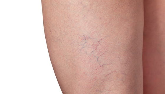
- Nodular GVMs look like dark purple or bluish bumps under the skin, creating a pebble-like appearance.
- Plaque-type GVMs are flat and can look like bruises.
- Solitary GVMs can grow larger, darker and more painful over time.
- GVMs most commonly form on:
- Fingers and toes
- Inside of the mouth
- Eyelids
- Some GVMs can be extensive and grow into the child’s arm or leg, involving the fat or muscle.
- The lesions can be painful, especially if they are bumped or pressed. For instance, a GVM on the sole of the foot could make it difficult for a child to walk.
- Very hot or cold temperatures can also cause GVMs to be painful.
What causes glomuvenous malformations?
GVMs are caused by a mutation (change) in a particular gene. In about 65% of cases, GVMs are inherited ― passed down from parent to child. GVMs in people within the same family may differ in severity ― some family members may have one lesion while others may have multiple GVMs.
The mutation can also arise on its own, causing GVMs in a child whose parents do not have the genetic mutation.
Glomuvenous Malformation Diagnosis
A doctor may notice a GVM under the child’s skin during physical examination. Further testing can rule out other venous malformations and conditions that look similar.
The best test to confirm a GVM is MRI, which can help the doctor visualize blood vessels and detect malformations. MRI can determine the exact location and extent of the malformed vessels and help the doctor plan a way to address them.
Glomuvenous Malformation Treatment
Not all GVMs require treatment, but a doctor may recommend medication or a procedure to remove a GVM if it is causing pain or growing larger or darker, which can happen over time.
There are several ways to treat GVMs:
- Medication with a drug called sirolimus can ease pain, soften the walls of the vessels and make GVMs appear lighter and less obvious.
- Sclerotherapy is a minimally invasive technique in which the doctor injects the GVM with a medication that gradually shrinks the lesion.
- Laser treatment can address GVMs close to the skin’s surface. A pulsed dye laser is designed to destroy the GVM without harming the surrounding area.
- Surgery may be able to safely remove small GVMs.
Living with Glomuvenous Malformations
Glomuvenous malformations are not associated with other problems, such as bleeding or cancer, and do not pose a risk to the child’s health. If symptoms are not severe, the impact on life is minimal.
However, when it comes to treatment, time is a factor. GVMs can become darker, larger and more difficult to treat as the child grows older. If you and your child’s doctor agree that treatment is preferred, the earlier therapy can take place, the better the results may be.


