Facial Paralysis in Children
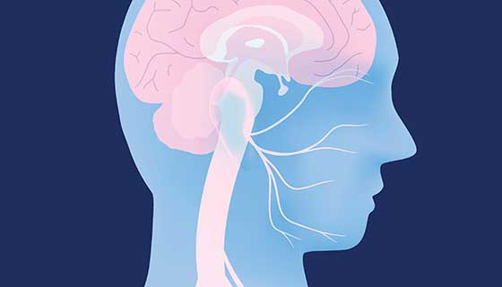
The seventh cranial nerve governs the sensation and movement of all the muscles in the face. Damage to this nerve can cause an inability to move one or both sides of the face, affecting a child’s ability to blink, speak, eat or convey emotion through facial expression.
What You Need to Know
- Facial paralysis in a child is rare, and it can be congenital (present at birth) or acquired.
- One or both sides of the child’s face may be affected.
- A majority of cases of facial paralysis in children resolve on their own, especially those resulting from a condition called Bell’s palsy.
- For facial paralysis that does not get better, it is important to see a specialist promptly for the best chance of a good outcome.
What causes facial paralysis in children?
Paralysis of a child’s facial muscles is a symptom with several possible underlying causes, including:
-
Bell’s palsy, which can be the result of viral infection or unknown cause
-
Trauma during birth
-
Head injury
-
Inborn conditions such as Moebius syndrome
-
Craniofacial abnormalities such as hemifacial microsomia
-
Tumors, including schwannomas or hemangiomas affecting the seventh cranial nerve
Symptoms of Facial Paralysis in Children
Symptoms may include
-
Noticeable drooping on one side of the face due to muscle weakness
-
Asymmetrical smile or facial expression
-
Inability to fully close eyes
-
Drooling
-
Difficulty feeding
-
Speech problems
Pediatric Facial Paralysis Diagnosis
In assessing a child with facial paralysis, the doctor will take a detailed history to determine when symptoms appeared, the severity of the paralysis and whether one or both sides of the child’s face are involved. The doctor may use a video camera to record the child’s range of movement.
The physician may recommend facial imaging with CT or MRI to point toward a diagnosis.
Treatment for Pediatric Facial Paralysis
Depending on the cause and severity of a child’s facial paralysis, nonsurgical therapies may be sufficient to treat the problem, including physical therapy and treatment with botulinum or steroid medication.
Facial Reanimation Surgery
Specialized surgical procedures can address severe or persistent facial paralysis in children, including these procedures:
-
Muscle transfers: The surgeon removes one or more tendons or muscles and relocates them to areas of the face where they can restore more natural movement. These procedures include:
-
Temporalis tendon transfer (also known as T3), which relocates one end of the temporalis tendon connected to the jaw and moves it closer toward the mouth. This procedure allows the child to smile by clenching their jaw. The T3 procedure takes about an hour, and may be performed in an outpatient setting.
-
Digastric tendon transfer, which relocates a tendon connected to a muscle located under the jaw.
-
Gracilis transfer, which transfers fibers from a slender muscle located in the inside of the thigh. This surgery may require multiple stages and a hospital stay of a few days, but it enables a more natural-looking and dynamic smile response that involves the entire face.
-
-
Nerve grafting involves moving nerves from different parts of the body to the face. Grafting can restore movement and increases muscle control.
Protecting the Child’s Eyes
Facial paralysis can affect a child’s ability to blink, resulting in dryness and potential damage to the eye. Your child may need to have their eye taped at night to protect it during sleep. One treatment your doctor may recommend is the attachment of a tiny platinum chain to the upper eyelid, which gently weighs the lid down and enables the child to blink and lubricate the eye with natural tears.


