Gastroparesis
Gastroparesis, also called gastric stasis, occurs when there is delayed gastric emptying. Delayed gastric emptying means the stomach takes too long to empty its contents. Sometimes, when the food doesn’t empty properly, it forms a solid mass called a bezoar. Although bezoars had magical powers in the Harry Potter books, usually these big masses of old food can block the stomach and lead to symptoms of nausea, vomiting and even obstruction of the stomach, which in turn may prevent food from passing into the small intestine.
Gastroparesis Symptoms
Gastroparesis often causes a number of nonspecific symptoms. It is important for a gastroenterologist to make a diagnosis. Symptoms of gastroparesis include:
- An early feeling of fullness
- Bloating
- Nausea
- Anorexia
- Vomiting
- Abdominal pain
- Weight loss
Gastroparesis Diagnosis
A diagnosis of gastroparesis begins with a comprehensive physical exam during which you describe your symptoms and medical history. Your doctor may find abdominal distention (swelling) or tenderness. Your doctor will also look for signs of underlying diseases or disorders that may be causing the gastroparesis. One big culprit can be diabetes. The high blood sugars from diabetes damage the nerves over time and can cause this delayed emptying. Treating the underlying cause will treat the gastroparesis as well.
During the physical exam, your doctor may listen for a "succession splash." The doctor will gently shake you and listen for the sound of fluid in your body. It can help confirm that there is an obstruction in your abdomen.
Your doctor may order laboratory tests as well. Although there is no definitive lab test for diagnosing gastroparesis, it may help rule out other underlying conditions.
Other tests your doctor may perform include:
- Endoscopy
- Upper gastrointestinal barium contrast radiography
- Gastric emptying scintigraphy
- Antroduodenal manometry
- Wireless motility study
Upper Endoscopy
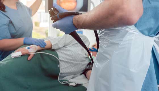
An endoscope is a thin, flexible tube that your doctor passes through your mouth and into your intestines. Upper endoscopy is performed using the endoscope in order to see the esophagus and stomach. Your doctor will most likely perform an upper endoscopy to rule out a mechanical obstruction at the outlet of the stomach, also called the pylorus. Obstruction is when there is a blockage of the intestines. The outlet of the stomach can have ulceration, damage or just a clog of food blocking the path. All of these can be seen at endoscopy.
During an upper endoscopy:
- You receive an anesthetic to help relax your gag reflex. You may also receive pain medication and a sedative.
- You lie on your left side, referred to as the left lateral position, and the endoscope is inserted through your mouth and pharynx and into the esophagus.
- The endoscope transmits an image of the esophagus, stomach and duodenum to a monitor that your physician is watching.
Upper Gastrointestinal Barium Contrast Radiography
Barium contrast radiography is an X-ray study. It is a commonly used procedure to diagnose gastroparesis.
During barium contrast radiography:
- You swallow a contrast solution called barium.
- The barium coats your esophagus and gastrointestinal tract, making it easier for the doctor to detect abnormalities.
- An X-ray is taken.
- Your doctor can determine if there are delays in the liquid emptying from your stomach.
Gastric Emptying Scintigraphy
Gastric emptying scintigraphy is the most commonly used test to confirm gastroparesis.
During the scintigraphy test, you will be given a special meal and sometimes a special drink. The meal will be radiolabeled with a marker that can be seen by the scanner. The goal of the test is to follow this special food as it travels through your system. Unlike diagnostic procedures that require you to fast beforehand, gastric emptying scintigraphy actually requires you to eat this special meal right before the test.
During a gastric emptying scintigraphy:
- You may be asked to have your blood sugar checked if you are diabetic. Blood sugar values over 250 can interfere with testing.
- You may be asked not to take narcotics for a time period before the test, as these medications can also interfere with the test.
- At Johns Hopkins, you are given a specially prepared meal of eggs, toast, jam and some water. Some centers provide other foods to be eaten. This meal has an exact number of calories and grams of fat, which helps us to compare the results of your test to those of other people.
- Prior to eating, your doctor will attach a radiotracer to the food. Don’t worry — the radiotracer has about as much radioactivity as a walk for an hour in a park when it’s sunny outside. It’s designed to be safe and well tolerated and to avoid causing harm.
- A scan is taken right away and then every hour after the meal is ingested for up to four hours. Your medical team will evaluate how the food you ingested moves through your stomach and gastrointestinal tract. A gastric emptying test may show a delay in emptying, which can help your doctors to make the diagnosis.
Antroduodenal Manometry
Antroduodenal manometry test the muscles used in digestion. During this procedure:
- Your doctor inserts a catheter through your mouth and into your stomach.
- The catheter stays in place for a period of six hours, in order to measure electrical and muscular activity in your stomach.
- You fast for the first few hours, so the doctor can record the measurements in a fasting state.
- Later, you eat a solid meal and your doctor records the measurements during digestion.
This procedure can help diagnose the exact cause of the gastroparesis. Doctors often use it for patients after standard treatments have failed, for surgery candidates and for patients with unexplained nausea.
Wireless Motility Study
A wireless motility study evaluates the time it takes for your stomach to empty. This is generally well tolerated and less invasive than other diagnostic studies.
During a wireless motility study:
- You ingest a small pill, which travels through your gastrointestinal system.
- As it travels, it collects data (temperature, acidity levels, pressure waves) and sends it to a data receiver that you wear, usually around your waist.
- The doctor sees the data, and you return to your doctor after a few days to review the results.
- The capsule is excreted in your bowel movements.
The advantage of this method is that you can continue with your normal activities while the capsule gathers the necessary information.


